Here is an explanation of the project, for the non-specialist. Below you will find more scientific details.
If you play a guitar string, you will hear a note, whose tone will depend on the string diameter and on the string tension. This sound is not just made of a pure (“single”) vibration, which would sound rather ugly and boring, but rather to the overlap of several acoustic frequencies playing simultaneously, which makes the note “round” and pleasant. More than that: it makes the note unique. You will be able, in fact, to tell it’s a guitar and not a piano, or even which kind of guitar. The very same concept applies to the voice of people: you can easily distinguish two persons pronouncing the same sentence at the phone because they have a different tone. The ensemble of the vibrating frequencies which form the sound and the voice is called the timbre. In physics, we call it spectrum, and it not only applies to sound but also to light, indicating its various frequency components, i.e. its colors!
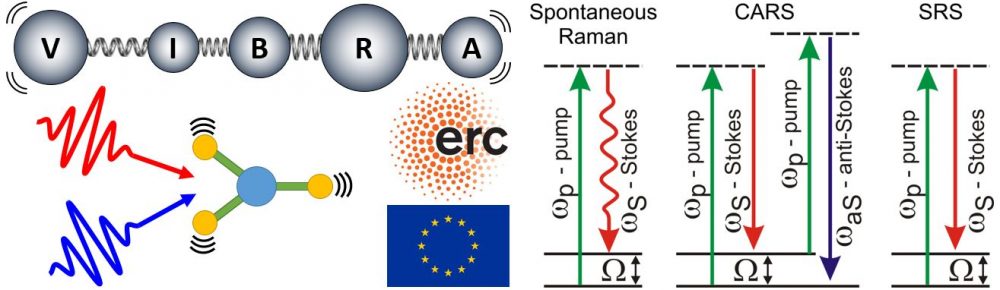
If you hit a molecule (which is a billionth time smaller than a guitar string) with a special hammer made of an extremely short light pulse, you can make it vibrate like a guitar string (but at a billion times higher frequency!). If you listen to the sound it makes (now you need a very special ultrasonic ear!), you can uniquely identify it and distinguish it from another molecule because it has a different oscillating pattern that we call vibrational spectrum. The basic concept, enabling very accurate chemical analysis of specimens, brings the name of its inventor Raman, Nobel laureate in Physics in 1930. Its recent implementation, using ultrashort laser pulses, is called coherent Raman process and enables higher sensitivity and thus higher acquisition speed, so that it is possible to apply it to microscopy. Coherent Raman Microscopy can visualize in real time the spatial distribution and concentration of several chemical species. Applied to biology, it can provide insights on the spatial arrangement of proteins, lipids, DNA, water and other constituents of cells. It is a so-called label-free technique, in the sense that it does not require any preparation of the sample by addition of fluorescent markers or other kind of stains, which could perturb the sample under examination or even contaminate it, altering the biological function. Furthermore, this technique does not require contact (it is stand-off) and it is nondestructive, because it employs infrared light, which is not absorbed by the sample, so that thermal load is minimized.
The European Research Council (https://erc.europa.eu/) has recently financed the Politecnico di Milano in Italy (www.polimi.it) to develop a next-generation microscopy system capable of rapidly visualizing in two dimensions the chemical content of a biological specimen using the coherent Raman process. If successful, the instrument will be applied to identify tumors in human biopsies with a higher accuracy and reproducibility than the actual practice (based upon the visual inspection of the tissue under a microscope and by the ensuing subjective opinion of a pathologist). In this way neoplasms can be located and their borders with normal tissue traced for surgery. This will pave the way towards future “virtual histopathology”: intraoperative non-invasive evaluation of cancerous tissue. The vision of this project is to allow researchers and doctors without a specific knowledge in lasers and optics to routinely visualize functional properties of cells and tissues in vivo.
In more detail: The VIBRA project aims at developing an innovative microscope for real-time non-invasive imaging of cells and tissues, which promises to have a revolutionary impact on several fields of biology and medicine. Chemically specific vibrational signatures of molecules enable their direct structural characterization. Reliable and quantitative endogenous bio-markers can be established, e.g., to follow cell differentiation and to identify crucial properties of tissues (malignant vs benign phenotype of a tumour). In this way neoplasms can be located and their borders with normal tissue traced for surgery.
Spontaneous Raman spectroscopy demonstrated this capability, but it is intrinsically too slow for imaging. Coherent Raman microscopy, on the other hand, can reach extremely high speed (up to the video rate) but at the expense of poor chemical selectivity, being limited to a single vibrational frequency.
The ground-breaking goal of VIBRA is to combine the most detailed molecular information over the entire vibrational spectrum with the highest acquisition speed. We will develop a complete coherent Raman microscope for near-video-rate broadband vibrational imaging. This high risk/high gain goal will be achieved by the combination of four key developments: improved pulsed laser source; optimized non-linear interaction, enhancing the signal; increase in acquisition speed, thanks to innovative spectrometers; parallel on-board data processing.
In the final application phase, the VIBRA project will validate the performances of the novel vibrational imaging system studying two important bio-medical problems: cancerous cell differentiation and detection of neuronal tumours. This will pave the way towards future “virtual histopathology”: intraoperative non-invasive evaluation of cancerous tissue. My vision is to allow researchers and doctors without a specific knowledge in lasers and optics to routinely visualize functional properties of cells and tissues in vivo.
This project has received funding from the European Research Council (ERC) under the European Union’s Horizon 2020 research and innovation programme (grant agreement No 648615”. ![]()