Raman microscopy provides functional information on tissues and cells. It is a non-contact, non-invasive and non-destructive method, as it does not require any sample labelling/staining. Every component of a biological specimen is characterized by a vibrational spectrum that reflects its molecular structure, such as this one:
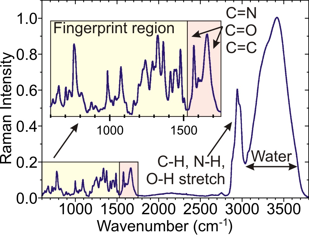
Typical Raman response of a cell. Adapted from J. R. Thomas, Annu. Rev. Biophys. Biomol. Struct. 28, 1 (1999), http://dx.doi.org/10.1146/annurev.biophys.28.1.1
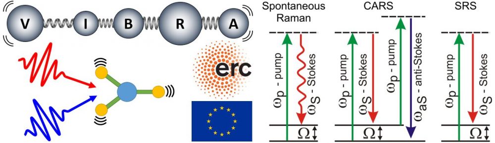
In spontaneous Raman microscopy a quasi-monochromatic laser at frequency ωp (“pump”) excites the molecules to a virtual state, which then relax to the ground state emitting photons with lower energy at frequency ωS (“Stokes”). The frequency shifts Ω=ωp-ωS match the molecular vibrations, which in turn reflect the molecular structure and become reliable and quantitative bio-markers to identify crucial properties/phenotypes of cells (differentiation, necrosis, cell cycle state…) and tissues (normal, cancerous or cultured tissues, collagen/elastin content…). The main drawback of spontaneous Raman is its very weak scattering cross section.
Coherent Raman Scattering (CRS) can overcome these hurdles, by setting up a coherent superposition of vibrational responses. When the frequency difference between two narrowband picosecond pulses (pump and Stokes) matches a characteristic vibrational frequency Ω, all the molecules are resonantly excited. This enhances the Raman signal by many orders of magnitude, enabling much higher imaging speeds. CRS microscopy provides further advantages: (i) being a nonlinear microscopy technique, the signal is generated only in the focal volume, thus allowing three-dimensional imaging; (ii) working out of resonance, it minimizes photo-damage to biological samples; (iii) compared to fluorescence microscopy, CRS is label-free and does not require sample labelling with exogenous fluorescent markers that often perturb the biological functions.
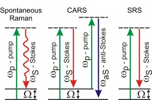
The two most widely employed CRS techniques are Coherent Antistokes Raman Scattering (CARS) and Stimulated Raman Scattering (SRS). CARS detects the coherent radiation at the anti-Stokes (blue-shifted) frequency ωaS = ωp + Ω. SRS monitors either the stimulated emission of the Stokes (called Stimulated Raman Gain, SRG) from a virtual state of the sample to the investigated vibrational state or the pump attenuation (called Stimulated Raman Loss, SRL).